
DIGITAL X-RAY MACHINE
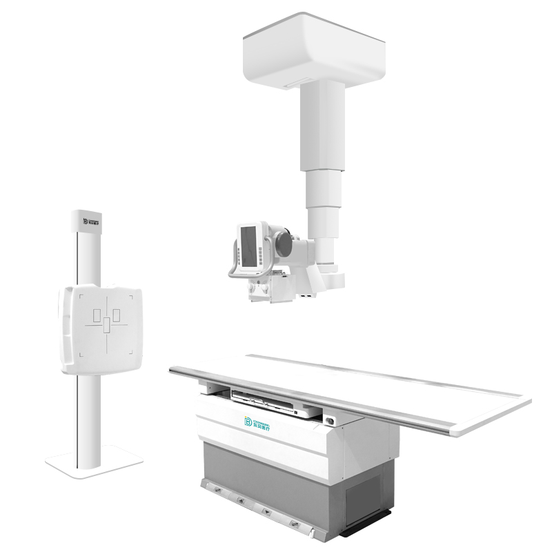

●Completely digitally configured DR
In radiology departments, which cope with heavy workload every day, doctors need a digitized medical X-ray imaging
system, which can present high-quality radiology images in a short period of time.
●Professional X-ray imaging system-a perfect assistant to doctors
Equipped with image processing software, the system, with reasonably categorized menuand visualized user interface,
is easy to use. By just clicking the mouse, doctors can choose which part of patients’ body is to be scanned. In addition
the system can set radiological parameters that should be applied to certain part of the body automatically, and quickly
present optimized radiology images.
●Exposure index and measurement display
A integrated exposure index and any exposure measurement display, measuring reference for operation technicians
and patients.
●Enhanced image sharpness and clarity
Although thickness of bones varies, advanced image processing technology effectively separates thick and thin parts
to display overlapping areas and contours more sharply and clearly.
●Ensured image integrity
Even in the presence of an implant, regions-of-interest (ROI) display clearly withourt artifacts. In areas where bones
overlap,each bone clearly displays.
●Unique AED acquisition technology
Configuration of the wireless tablet can seamlessly with hospital other X-ray machine, no need to change any line. To many hospital equipment upgrades for DR digital equipment.
●Excellent dynamic range
Because the regions-of-interest (ROI) contrast is improved, a clear, detailed display of bone and soft tissue in a single
image is provided. Patients’ organs such as lung, spine and inguinal region with different characteristics can be
displayed in detail on a single page.
|
Category
|
Parameters
|
|
Output Power
|
65.5kW
|
|
Dual-focus
|
0.6/1.2mm
|
|
Inverter Frequency
|
110kHz
|
|
Continuous Fluoroscopy
|
Tube Voltage: 40-150kV
|
|
Tube Current:10-800mA
|
